

WHAT IS OESOPHAGEAL ATRESIA?
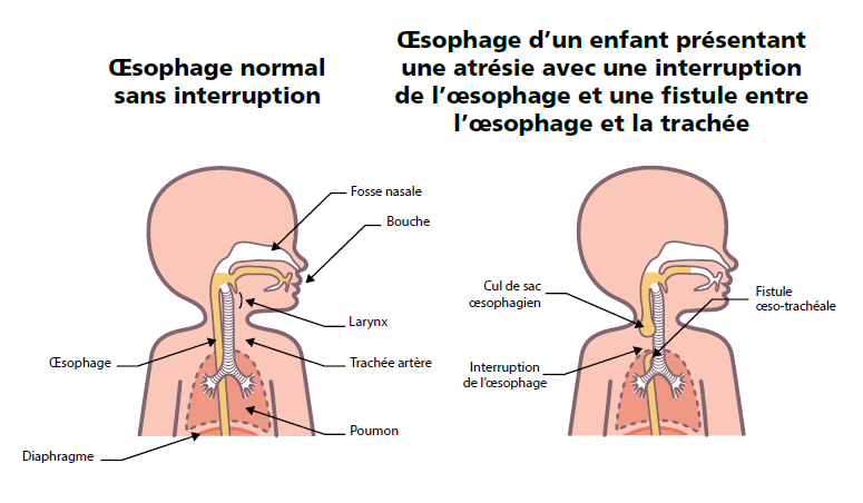
Oesophageal atresia is a malformation of the oesophagus (the “tube” connecting the mouth to the stomach) that is present at birth. It is characterised by an interruption in the continuity of the oesophagus, which thus ends in a “cul-de-sac”: food or saliva therefore cannot reach the stomach.
Atresia is most often associated with an abnormal connection between the oesophagus and the trachea (the duct that brings air to the lungs). This abnormal connection is called a tracheo-oesophageal fistula. In case of fistula, food can pass into the trachea and thus into the lungs, which can lead to serious respiratory problems. These anomalies can be corrected surgically.
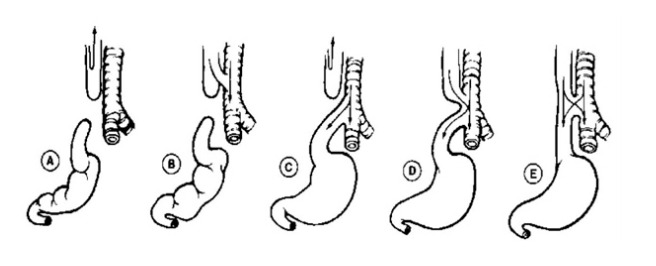
There are five types of oesophageal atresia defined as a function of the fistula(s):
- type-I (or A) atresia: isolated atresia without fistula;
- type-II (or B) atresia atresia with fistula in the upper segment of the oesophagus;
- type-III (or C) atresia atresia with fistula in the lower segment of the oesophagus;
- type-IV (or D) atresia variant of type-III atresia;
- type-V (or E) atresia atresia with tow or more fistulas.
The most common form is type-III (or C) atresia.
Approximately one child in 3,000 is born with oesophageal atresia with or without fistula. This malformation affects girls and boys equally.
HOW IS THIS MALFORMATION DIAGNOSED?
Oesophageal atresia may be suspected prior to birth if hydramnios or evocative signs by ultrasound are found. However, diagnosis is generally done after birth:
- in case of excessive salivation or coughing by inhalation or during feeding, in case of regurgitation of undigested milk or swallowing difficulties with inhalation into the lungs;
- by passage of an oesophageal probe, which is stopped around the upper cul-de-sac.
To visualise the atresia and to determine its type and the location of any tracheo-oesophageal fistula, an X-ray of the abdomen is done quickly.
WHAT IS THE TREATMENT?
Treatment of oesophageal atresia consists in surgery to close the tracheo-oesophageal fistula and to connect the two parts of the oesophagus together.
Before the oesophagus is “repaired”, the baby cannot be fed normally. Some children must be fed via gastrostomy, especially if they are premature and cannot be operated on quickly. Gastrostomy involves inserting a small plastic tube directly connecting the stomach to the outer wall of the belly. This makes it possible to introduce liquid foods directly into the stomach (enteral nutrition). It is removed as soon as the baby is able to feed via the mouth.
Sources
Learn more:
-
PNDS (French national diagnosis and care protocol)
-
Guidelines "Évaluation et le traitement des complications gastro-intestinales et nutritionnelles chez l'enfant atteint d'atrésie de l’œsophage et de fistule oeso-trachéale" (ESPGHAN-NASPGHAN)
-
Position Paper of INoEA Working Group on Long-Gap Esophageal Atresia : For Better Care
Relevant reference centre:
Patients association